Amblyopia Preferred Practice Pattern®


Amblyopia Preferred Practice Pattern®

Created
Tags Pediatric
Journal Ophthalmology
Status 審查完成
校稿者 蕭靜熹 醫師

中文摘要

本文探討了青光眼、斜視、結合性弱視等因素引起的弱視治療,以及少數與眼睛或視覺途徑結構異常相關的視覺剝奪性弱視。此外,本文提到,有特定的視覺要求,包括立體視,適用於多種職業,如軍事、执法、航空和手術。但目前尚缺乏證據表明單眼弱視會影響教育或職業表現。最後,本文提到,弱視是一種需要醫學治療的醫療狀況,醫療保險計劃應覆蓋所有類型的弱視治療。

English Abstract

The article discusses the treatment of amblyopia caused by factors such as glaucoma, strabismus, and combined amblyopia, as well as visual deprivation amblyopia associated with structural abnormalities involving the eye or visual pathway. Additionally, the article mentions that there are specific visual requirements, including stereopsis, for various career fields such as the military, law enforcement, aviation, and surgery. However, there is currently insufficient evidence that unilateral amblyopia is an impediment to education or career performance. Finally, the article notes that amblyopia is a medical condition that requires medical treatment, and healthcare insurance plans should cover all types of amblyopia management, including timely screening, treatment, and monitoring for recurrence.

HIGHLIGHTED FINDINGS AND RECOMMENDATIONS FOR CARE

Treatment of refractive error alone can improve visual acuity (VA) in children who have anisometropic, strabismic, or combined amblyopia. Visual acuity of children who have bilateral refractive amblyopia also can substantially improve with refractive correction alone.

Most children who have moderate amblyopia (20/40 to 20/80) respond to initial treatment consisting of 2
hours of daily patching or weekend atropine.

Following treatment of amblyopia caused by strabismus, anisometropia, or both, continued monitoring is necessary and additional treatment, if needed, is associated with long-term durability of the VA improvement.


Suitable treatment options for amblyopia include optical correction, patching, pharmacological treatment, optical treatment, Bangerter (translucent) filters, and digital therapeutics, in addition to managing the underlying cause of amblyopia.


Amblyopia treatment may be effective in older children and adolescents, particularly if they have not previously been treated.

Esotropia and Exotropia Preferred Practice Pattern®


Esotropia and Exotropia Preferred Practice Pattern®

Created
Tags Pediatric
Journal Ophthalmology
Status 審查完成
校稿者 蕭靜熹 醫師

中文摘要

眼科的Preferred Practice Pattern®指南強調對斜視和弱視患者進行及時和適當的治療的重要性。在一些情況下,兒童的斜視可能會在沒有治療的情況下自行消失,而在其他情況下,可能需要進行手術干預。像Worth 4-Dot Testing和立體視力測試等評估應該進行,以評估兒童的雙眼狀態。Retinoscopy需要充分的睫狀肌麻痺,視神經檢查對於檢測水腫、蒼白或先天異常等徵象至關重要。

如果眼鏡和弱視治療無法緩解內斜視,應考慮進行手術矯正。對於幼兒內斜視,早期手術矯正可能會改善立體感。對於間歇性外斜視的情況,應先採用保守治療方式。過度矯正的凹透鏡(overcorrecting minus-lens)治療雖然有效,但結果通常是暫時性的,並可能導致近視的加劇。定期的追蹤和全面的眼科檢查對於監測患者的進展並確保在需要時提供適當的屈光幫助非常重要。

English Abstract

The Preferred Practice Pattern® guidelines for eye care emphasize the importance of timely and appropriate treatments for patients with strabismus and amblyopia. In some cases, strabismus in children may resolve without treatment, while in others, surgical intervention may be necessary. Assessments like the Worth 4-Dot Testing and stereoacuity tests should be performed to evaluate a child’s binocular sensory status. Accurate retinoscopy in children requires adequate cycloplegia, and optic nerve evaluations are crucial for detecting signs of swelling, pallor, or congenital anomalies.

Surgical correction should be considered for children with esotropia if eyeglasses and amblyopia management prove insufficient. For infantile esotropia, early surgical alignment may improve stereoacuity outcomes. In cases of intermittent exotropia, conservative methods should be employed before surgery. Overcorrecting minus-lens therapy has shown effectiveness, but results are often temporary and may lead to a myopic shift. Regular follow-ups and comprehensive ophthalmic examinations are essential for monitoring patients’ progress and ensuring proper refractive aids are provided when needed. Further studies are necessary to assess the long-term impact of these interventions across diverse populations.

HIGHLIGHTED FINDINGS & RECOMMENDATIONS FOR CARE


Strabismus in children under 4 months of age sometimes resolves without treatment, particularly if the deviation is intermittent, variable, or measures less than 40 prism diopters.


Repeat cycloplegic refraction is indicated when esotropia does not respond to an initial prescription for hyperopia or when esotropia recurs after surgery.


Acquired esotropia should be evaluated and treated promptly.


Young children with intermittent exotropia and good fusional control can be followed without surgery because there is a low rate of deterioration to constant exotropia or reduced stereopsis.


Indications for surgery in intermittent exotropia include a progression to constant or nearly constant deviation, reduced stereopsis, and/or a negative effect on social interactions.


Unilateral recess-resect and bilateral lateral rectus recessions are both effective initial surgical procedures for the treatment of intermittent exotropia.


Convergence insufficiency occurs in children and adults, and symptoms with near viewing can often be improved using vergence exercises.


Ophthalmic Technology Assessment: Surgical Treatments to Improve Visual Acuity in Infantile Nystagmus Syndrome


Ophthalmic Technology Assessment: Surgical Treatments to Improve Visual Acuity in Infantile Nystagmus Syndrome

Created
Tags Pediatric
Journal Ophthalmology
Status 審查完成
校稿者 蕭靜熹 醫師

Ophthalmology Volume 130, Number 3, March 2023

中文摘要

本文回顧了有關治療幼年期眼震症候群 (Infantile Nystagmus Syndrome, INS) 的手術方式的文獻。研究共有23篇,其中包括1篇隨機試驗和22篇病例系列。目前沒有一種標準的手術治療INS以改善最佳矯正視力,但一些手術方法,如large recessions, anterior extirpation, myectomy with or without pulley fixation, and transposition of extraocular muscles,可以改善視力和減緩眼震症狀。研究顯示,幼年期手術的療效可能比成年期更佳。未來的研究需要探討新的手術技術和替代療法。此外,除了應該使用標準的光學字符進行雙眼視力測試外,還應包括客觀的眼震圖學(nystagmography)作為次要結果評估。最後,需要進行一項前瞻性、隨機、盲性研究,以確定手術治療是否有益。

English Abstract

The article provides a summary of various surgical procedures to improve visual acuity in patients with Infantile Nystagmus Syndrome (INS). The authors reviewed 23 studies, including one randomized trial and 22 case series, that evaluated surgical options for INS. The surgical procedures included large recessions, anterior extirpation, myectomy with or without pulley fixation, and transposition of extraocular muscles. The studies reported improvements in binocular visual acuity, ranging from 0.1 to 0.3 logMAR, or 1 to 3 Snellen lines, with statistical significance reported in some cases. However, the clinical significance of these improvements is debatable. There was no clear difference among the various procedures in terms of visual acuity outcomes. Objective nystagmography should be included as a secondary outcome measure along with visual acuity testing. Additional research is necessary to validate methods of VA testing in nonverbal children with INS.

Refractive Surgery Preferred Practice Pattern®

Refractive Surgery Preferred Practice Pattern®

Created
Tags CGMHOPH Cornea Refraction
Journal Ophthalmology
Status 審查完成
校稿者 蕭靜熹 醫師

Secretary for Quality of Care
Timothy W. Olsen, MD
Academy Staff
Andre Ambrus, MLIS
Meghan Daly
Flora C. Lum, MD
Medical Editor: Susan Garratt
Approved by: Board of Trustees
September 9, 2022
Copyright © 2022 American Academy of Ophthalmology®

English Summary :

Keratorefractive surgery can be used for a wide range of refractive errors, but intraocular procedures may be considered in some cases. A comprehensive medical eye evaluation should be performed before any refractive surgery procedure. Excimer laser ablation using wavefront aberrometry information can limit the induction of higher-order aberrations (HOAs) and even reduce pre-existing HOAs in some cases. The most common complaints from patients dissatisfied with refractive surgery include blurred vision, glare, dry eyes, and night-vision problems. Intraocular refractive surgery can be considered for patients who wish to reduce their dependence on eyeglasses or contact lenses. Potential complications of refractive lens exchange for myopia include retinal detachment, infectious endophthalmitis, intraoperative suprachoroidal hemorrhage, cystoid macular edema, corneal edema, and intraocular lens dislocation.

中文摘要:

角膜屈光手術可用於廣泛的屈光不正,但在某些情況下,可能會考慮使用眼內手術。在進行任何屈光手術之前,應對患者進行全面的眼科檢查評估。使用Excimer laser ablation using wavefront aberraometry可以限制高階像差(HOAs)的產生,並在某些情況下減少現有的HOAs。對屈光手術不滿意的患者最常見的抱怨包括視力模糊、眩光、眼睛乾燥和夜間視力問題。對於希望減少對眼鏡或隱形眼鏡依賴的患者,可以考慮進行眼內屈光手術。近視屈光晶狀體置換術的潛在併發症包括視網膜脫離、感染性眼內炎、手術中超脈絡膜出血、囊樣黃斑水腫、角膜水腫和人工晶狀體脫位。

HIGHLIGHTED FINDINGS AND RECOMMENDATIONS FOR CARE

Contraindications to refractive surgery include the following:

  • Unstable refraction
  • Abnormalities of the cornea (e.g., keratoconus or other corneal ectasias, thinning, edema, interstitial or neurotrophic keratitis, extensive vascularization)
  • Insufficient corneal thickness for the proposed ablation depth
  • Visually significant cataract
  • Uncontrolled glaucoma
  • Uncontrolled external disease (e.g., blepharitis, dry eye syndrome, atopy/allergy)
  • Uncontrolled autoimmune or other immune-mediated disease
  • Uncontrolled mental illness, including anxiety or depression
  • Unrealistic patient expectations

It is recommended that corneal refractive surgery patients be provided with a record listing diagnosis, preoperative keratometry readings, and refraction, as well as postoperative refraction.


It is recommended that the refractive surgeon maintain a record including preoperative keratometry and refraction as well as postoperative refraction and provide that data if needed for future eye care, including cataract surgery.


As part of the informed consent process, it is recommended that the refractive surgeon review common adverse effects such as dry eye and eventual presbyopia with patients considering corneal refractive surgery.


Excimer laser ablations that result in very thin residual stroma increase the risk for ectasia. For laser in situ keratomileusis (LASIK) procedures, a minimum of 250 µm is suggested as a safe residual stromal bed thickness. There is no absolute value that guarantees ectasia will not occur. Abnormal topography and percentage of tissue altered (PTA) higher or equal to 40% are also associated with higher ectasia risk.


Published studies have failed to demonstrate a relationship between pupil size and the quality of postop vision, minimizing the importance of pupillometry in the preoperative workup.
Persistent diffuse lamellar keratitis (DLK) unresponsive to corticosteroids should prompt consideration of microbial keratitis or interlamellar fluid due to increased intraocular pressure (IOP) measured peripheral to the LASIK flap, intraocular inflammation, or endothelial decompensation. For extensive DLK, the interface should be irrigated to minimize stromal loss and changes in refractive correction.


Surgical management of presbyopia includes keratorefractive surgery, corneal inlays, and intraocular lens implantation (multifocal, accommodative, and extended depth of focus lenses).

Refractive Errors Preferred Practice Pattern®

Refractive Errors Preferred Practice Pattern®

Created
Tags CGMHOPH Pediatric Refraction6
Journal Ophthalmology
Status 審查完成
校稿者 蕭靜熹 醫師

中文摘要

本文介紹了眼球屈光不正的相關問題及其治療方法。
眼球屈光不正是一種常見的視覺問題,包括近視、遠視和散光。
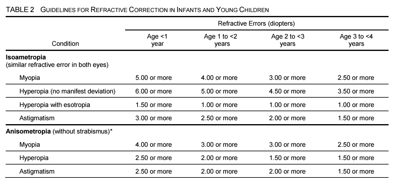
視力檢查和眼科檢查對於確定屈光不正的診斷和治療方案至關重要。矯正視力是診斷和治療眼球屈光不正的首要方法,以提高視力和減少不適感。選擇適合的眼鏡鏡框和鏡片對於矯正視力非常重要。
隱形眼鏡是另一種矯正視力的方法。在隱形眼鏡的選擇方面,應考慮患者的生活方式和需要。在安全和舒適性方面,目前的隱形眼鏡比過去有很大的改進。在選擇隱形眼鏡之前,應該進行完整的眼科檢查,以確定是否適合使用隱形眼鏡,以及適合哪種類型的隱形眼鏡。
在隱形眼鏡的使用中,適當的保養和清潔非常重要。不遵從隱形眼鏡的建議使用方法和保養方法,可能會導致角膜損傷和感染。在使用隱形眼鏡時,患者應注意眼睛是否有不適感,如灼熱、疼痛、視力模糊、紅斑和分泌物等。
有關角膜炎的研究顯示,Pseudomonas為使用隱形眼鏡相關的角膜炎最常見的病原體,而兒童的微生物性角膜炎風險相對較高。角膜炎風險增加的原因包括使用同一隱形眼鏡超過規定期限、隱形眼鏡清潔不當以及睡覺時未取出隱形眼鏡。在診斷和管理隱形眼鏡相關的問題時,醫護人員需要進行全面的眼科評估,包括評估患者的角膜和眼表,並詢問患者有無相關問題,例如眼睛紅腫、疼痛或異物感。
另一方面,鑑於許多人需要眼鏡或隱形眼鏡進行視力矯正,建議對於眼科醫生和患者,掌握當前的矯正方法以及各種矯正方法的適應症和禁忌症。
眼科醫生需要對不同人群的矯正方式進行全面的評估,包括患者的年齡、病史、角膜形態、視力問題的嚴重程度以及患者的職業和活動需求。例如,對於青少年和兒童,尤其是那些有近視風險的人,可以採用對抗近視進展的治療方法,如使用矯正光學和配戴角膜塑形鏡片等。
對於老年人或其他需要視力矯正的人,可以考慮使用多焦點鏡片或漸進式眼镜等多種矯正方法,以滿足他們不同距離的視力需求。此外,RGP lenses 是治療高度近視和高度散光的有效方法。需要指出的是,醫生在選擇矯正方法時應注意患者的眼睛健康和安全,以避免出現相關併發症。

English Abstract

This article discusses the related issues and treatment methods for refractive errors of the eye. Refractive errors of the eye are a common vision problem, including myopia, hyperopia, and astigmatism.

Vision and eye examinations are essential for determining the diagnosis and treatment plan for refractive errors. Correcting vision is the primary method for diagnosing and treating refractive errors of the eye, to improve vision and reduce discomfort. Choosing the appropriate eyeglass frames and lenses is critical for correcting vision.

Contact lenses are another method for correcting vision. In selecting contact lenses, patients’ lifestyles and needs should be considered. In terms of safety and comfort, current contact lenses have greatly improved compared to the past. Before selecting contact lenses, a complete eye examination should be conducted to determine if contact lenses are suitable and which type of contact lenses are appropriate.

Proper maintenance and cleaning are essential for contact lens use. Failure to follow the recommended usage and maintenance methods for contact lenses may lead to corneal damage and infection. When using contact lenses, patients should pay attention to any discomfort, such as burning, pain, blurred vision, redness, and discharge.

Research on keratitis shows that Pseudomonas is the most common pathogen associated with contact lens-related keratitis, and children are relatively more at risk for microbial keratitis. The reasons for increased risk of keratitis include using the same contact lenses beyond the prescribed time frame, improper contact lens cleaning, and failure to remove contact lenses while sleeping. When diagnosing and managing contact lens-related problems, healthcare professionals need to conduct a comprehensive eye assessment, including evaluating the patient’s cornea and ocular surface, and inquire about any relevant issues such as eye swelling, pain, or foreign body sensation.

On the other hand, considering that many people require glasses or contact lenses for vision correction, it is recommended for ophthalmologists and patients to be familiar with the current correction methods and the indications and contraindications for various correction methods. Ophthalmologists need to conduct a comprehensive evaluation of the correction methods for different populations, including the patient’s age, medical history, corneal morphology, severity of visual problems, and the patient’s occupation and activity needs. For example, for adolescents and children, especially those at risk for myopia, treatment methods to counteract myopia progression, such as using correcting optics, and wearing orthokeratology lenses, can be used.

For elderly individuals or others who require vision correction, various correction methods such as multifocal lenses or progressive eyeglasses can be considered to meet their visual needs at different distances. Additionally, rigid gas-permeable contact lenses are an effective method for treating highly myopic and highly astigmatic conditions. It should be noted that doctors should pay attention to patients’ eye health and safety when selecting correction methods to avoid related complications.

HIGHLIGHTED FINDINGS AND RECOMMENDATIONS FOR CARE

The prevalence of myopia is increasing in the United States and other industrialized societies. Increased time spent outdoors appears to be protective against myopia in children. Increased levels of near work are less of a risk factor than previously believed.


Increased outdoor time and low-concentration atropine have been shown to reduce the likelihood of myopia onset.


Antimuscarinic eyedrops, multifocal spectacles and contact lenses, and overnight orthokeratology have been shown to be varibly effective in some populations for myopia control, that is, to reduce the progression of myopia in school age children.


Studies from around the world have confirmed that that the incidence of microbial keratitis has not been reduced with the introduction of new lens types and that overnight wear of any contact lens is associated with a higher risk of infection than daily wear.


Although there are lenses approved by the FDA for extended wear, alternatives should be presented to patients for whom this mode of contact lens wear is being considered because overnight wear, regardless of contact lens type, increases risk of microbial keratitis.


Daily disposable contact lenses (rather than planned replacement lenses) are the safest lenses with the lowest rate of complications associated with soft contact lens wear. No-rub cleaning, topping off (reuse) of solutions, contaminated lens cases, exposure to tap water, wearing contact lenses in hot tubs and showers and while swimming, and changes in water supply are associated with Acanthamoeba and fungal keratitis related to contact lens wear in the recent decades.


Hydrogen peroxide systems are superior to multipurpose solutions for reducing the likelihood of infections or inflammatory complications; they are the preferred mode of nightly disinfection for patients who cannot wear daily disposable lenses, especially if they have had complications of contact lens wear in the past.


Presbyopia can be managed by using eyeglasses; contact lenses; topical agents; intraocular lenses with multifocal, accommodating, or extended depth of focus features; and monovision strategies with contact lenses or intraocular lenses. Adverse events related to FDA-approved drugs and devices should be reported to MedWatch
(www.fda.gov/medwatch)

Pediatric Eye Evaluations Preferred Practice Pattern®

Pediatric Eye Evaluations Preferred Practice Pattern®

Created
Tags CGMHOPH Pediatric Refraction
Journal Ophthalmology
Status 審查完成
校稿者 蕭靜熹 醫師

中文摘要

視力篩查通常在兒科診所由護士或其他受過培訓的醫護人員進行。兒童視力障礙和失明的原因在全球各地差異很大。在低收入國家,白內障和由傳染病或維生素A缺乏引起的角膜病變是導致嚴重視力障礙和失明的常見原因。

兒童應在整個童年和青少年時期接受定期視力篩查,因為隨著孩子的成長,可能會出現視力問題。大多數基於儀器的視力篩查方法可以檢測弱視的風險因素,如斜視、高度或不對稱的屈光不正、晶體混濁(如白內障)、視網膜異常(如視網膜母細胞瘤)和眼瞼下垂。在社區中,照相篩檢設備對弱視風險因素評估有高的敏感性和特異性。

執行視力篩查的醫生、護士、眼科醫生、驗光師、視光師、教師和其他執業人員應接受培訓,以便評估視力問題的風險因素、檢測眼睛結構性的問題、評估不同年齡兒童的視力,並/或進行儀器篩查。對於無法進行常規視力測試的兒童,應在6個月內(如有可能,更早)重新篩查或接受全面眼科檢查。

總之,對兒童和青少年進行定期視力篩查非常重要。

English Abstract

Vision screening is typically conducted in pediatric clinics by nurses or other trained healthcare professionals. The causes of severe vision impairment and blindness in children vary considerably worldwide, with cataracts and corneal conditions due to infectious diseases or vitamin A deficiency being common causes in low-income countries.

Children should undergo periodic vision screenings throughout childhood and adolescence, as vision problems may arise as they grow. Most instrument-based vision screening methods detect risk factors for amblyopia, such as strabismus, high or asymmetric refractive errors, media opacities (e.g., cataract), retinal abnormalities (e.g., retinoblastoma), and ptosis. In community settings, photoscreening devices demonstrate reasonably high sensitivity and specificity for amblyopia risk factors.

Physicians, nurses, ophthalmologists, optometrists, orthoptists, teachers, and other professionals performing vision screening should be trained to assess risk factors for vision problems, detect structural eye issues, evaluate visual abilities in children of different ages, and/or conduct instrument-based screenings. Children who are untestable should be rescreened within six months (preferably sooner) or referred for a comprehensive eye examination.

In summary, regular vision screenings for children and adolescents are essential in identifying and addressing potential vision problems to ensure healthy visual development.

HIGHLIGHTED RECOMMENDATIONS FOR CARE

Amblyopia meets the World Health Organization criteria for a disease that benefits from screening because it is an important health problem for which there is an accepted treatment, it has a recognizable latent or early symptomatic stage, and a suitable test or examination is available to diagnose it before permanent vision loss
occurs. The U.S. Preventive Services Task Force (USPSTF) recommends vision screening at least once for all children aged 3 to 5 years to detect amblyopia or its risk factors.

4 Vision testing with single optotypes is likely to overestimate visual acuity in a patient who has amblyopia. A more accurate assessment of monocular visual acuity is obtained by presenting a line of optotypes or a single optotype with crowding bars that surround (or crowd) the optotype being identified.
The choice and arrangement of optotypes (letters, numbers, symbols) on an eye chart can significantly affect the visual acuity score obtained. The preferred optotypes are LEA symbols, HOTV, and Sloan letters because they are standardized and validated.5, 6 Instrument-based screening techniques, such as photo screening and autorefraction, are useful for assessing amblyopia and reduced-vision risk factors for children ages 1 to 5 years, as this is a critical time for visual development.7 Instrument-based screening can also be used for older children who are unable to participate
in optotype-based screening. This type of screening has been shown to be useful in detecting amblyopia risk factors in children with developmental disabilities.8
Vision screening should be performed at an early age and at regular intervals throughout childhood to detect amblyopia risk factors and refractive errors. The elements of vision screening vary depending on the age and level of cooperation of the child, as shown in Table 1.

[/av_textblock]

Surgical Results of Trabeculectomy among Groups Stratified by Prostaglandin Associated Periorbitopathy Severity

Surgical Results of Trabeculectomy among Groups Stratified by Prostaglandin Associated Periorbitopathy Severity

Created
Tags Glaucoma
Journal Ophthalmology
Status 審查完成
校稿者 蕭靜熹 醫師

Ophthalmology Volume 130, Number 3, March 2023

中文摘要

本篇研究的主要目的是探討病人使用Shimane University prostaglandin-associated periorbitopathy
syndrome Grading System (SU-PAP)分級系統,其和青光眼手術(Trabeculectomy)的成效之間是否存在相關性。研究對象為有原發性開放性青光眼、未接受結膜切開手術、完成所有12個月後續追蹤,並利用SU-PAP系統評估其青光眼手術前的病情嚴重程度,共計收集了兩家醫院205例病例,最終納入139位日本患者的眼睛進行分析。研究顯示,手術成果與年齡、性別、手術前眼壓和藥物治療、屈光狀態和是否同時進行白內障手術等因素無關,但是SU-PAP分級系統和手術成效有關。愈厲害的SU-PAP病人的手術成功率較低。研究結果顯示,在進行青光眼手術時,應留意患者的PAP程度,並根據其情況選擇相應治療方式。

English Abstract

The study aimed to evaluate the surgical success rates of trabeculectomy among groups stratified by prostaglandin-associated periorbitopathy (PAP) severity. The Shimane University PAP Grading System (SU-PAP) was used to grade PAP severity among 139 consecutive eyes of 139 Japanese subjects with primary open-angle glaucoma who underwent trabeculectomy between June 2018 and December 2020. The study found that higher SU-PAP grades were associated with lower success rates for intraocular pressure control. Needling, bleb revision, and surgery other than glaucoma were more frequent in patients with higher SU-PAP grades. However, age, gender, preoperative intraocular pressure and medications, refractive error, and simultaneous cataract surgery were not associated with surgical failure. The study suggests that to retain surgical effectiveness, physicians should prevent patients from progressing to severe PAP, an avoidable side effect, by switching or stopping the causative medications. The study also recommends that treating physicians pay attention to patients with severe PAP because it can affect glaucoma management. However, the study acknowledges limitations due to its retrospective design and the potential impact of other glaucoma medications on the results. In conclusion, the study highlights the importance of considering PAP severity when performing trabeculectomy and managing glaucoma.

附註 The SU-PAP grading system classifies the severities of PAP into 4 grades based on the appearance and difficulty performing Goldmann applanation tonometry (GAT).

Grade 0 (no PAP): no prostaglandin-associated cosmetic changes by macroscopic or slit-lamp observation

Grade 1 (superficial cosmetic PAP): the presence of eyelid hyperpigmentation or eyelash growth

Grade 2 (deep cosmetic PAP): the presence of at least 1 of DUES, blepharochalasis
involution, periorbital fat loss, and enophthalmos

Grade 3 (tonometric PAP): difficulty performing GAT or reduced reliability of GAT due to the presence of PAP-related deepening of the upper eyelid sulcus, hardening of eyelids, ptosis, or enophthalmos.

Randomized Trial to Evaluate the Efficacy of the Nanodropper Device for Pupillary Dilation and Cycloplegia in Children

Randomized Trial to Evaluate the Efficacy of the Nanodropper Device for Pupillary Dilation and Cycloplegia in Children

Created
Tags CGMHOPH Pediatric,Refraction
Journal Ophthalmology
Status 審查完成
校稿者 蕭靜熹 醫師

Ophthalmology Volume 130, Number 3, March 2023

中文摘要

本研究旨在評估一種新型小容量滴眼器套件「Nanodropper」對於幼童瞳孔擴張和消除調節反應的有效性。本研究將患者分為兩組,隨機將小容量擴瞳滴眼液和標準滴眼液分別注入兩只眼睛,之後進行檢查,並以三個指標評估治療效果:spherical equivalent、瞳孔收縮百分比和散瞳後最大瞳孔直徑。結果顯示,使用「Nanodropper」治療組的最大瞳孔直徑達到非劣效,但瞳孔收縮百分比和spherical equivalent等效度在統計上沒有達到非劣效,但無臨床意義。本研究認為,小容量滴眼液具有減少不必要的醫療浪費和藥物毒性的潛力,且有效性與標準滴眼液相近。

English Abstract

The article discusses a study conducted at the Pediatric Ophthalmology Clinic of the University of California, San Francisco, which aimed to evaluate the effectiveness of small-volume eye drops for pediatric pupillary dilation and cycloplegia using the Nanodropper. Participants were randomized to receive small-volume dilating drops in one eye and standard of care dilating drops in the other eye. A non-inferiority analysis was performed to assess changes from before to after dilation in spherical equivalent, pupil constriction percentage, and maximum pupil diameter after dilation. The Nanodropper achieved non-inferiority for maximum pupil diameter after dilation, but not for spherical equivalent or constriction percentage. However, the small differences in the effect of the Nanodropper versus standard of care on all primary outcomes were not clinically significant. The study concludes that small-volume eye drops have the potential to decrease unnecessary medical waste and medication toxicity while maintaining therapeutic effect. The article notes that larger eye-drop volumes may increase the likelihood of adverse medication effects, especially because topical medications that reach the nasolacrimal duct and nasopharynx can be absorbed systemically through the mucosa, avoiding first-pass metabolism. The study demonstrates that small-volume eye drops are similar to standard of care eye drops for pupillary dilation and cycloplegia in pediatric patients. Using smaller-volume eye drops has economic and health care system advantages, as it could result in cost savings for patients, health care providers, and the medical system.

Longitudinal Changes and Predictive Value of Choroidal Thickness for Myopia Control after Repeated Low-Level Red-Light Therapy

3Longitudinal Changes and Predictive Value of Choroidal Thickness for Myopia Control after Repeated Low-Level Red-Light Therapy

Created
Tags CGMHOPH Pediatri,,Refraction
Journal Ophthalmology
Status 審查完成
校稿者 蕭靜熹 醫師

中文摘要

本篇研究報告指出,經過repeated low-level red-light (RLRL)治療1年後,近視兒童的視網膜脈絡膜厚度(mCT)有所改變,且此改變具有預測對於控制近視的治療效果的價值。該研究採用隨機對照試驗,比較RLRL與單視覺鏡片對於控制近視的效果,結果顯示RLRL治療相對於single-vision spectacle(單視覺鏡片)使用能夠減少69.4% axial elongation 和 76.6% spherical equivalent refraction progression。此外,該研究還證實RLRL治療可以促進mCT的增厚,這表明脈絡膜增加的血流可能是對控制近視效果的機制。然而,即使在至少75%的治療遵從度下,脈絡膜仍會變薄,原因仍需進一步研究。該研究建議,只需檢測3個月的mCT變化即可預測12個月的近視控制效果,並呼籲未來的研究應該進一步探討RLRL治療後脈絡膜增厚的機制。

English Abstract

This study aimed to evaluate longitudinal changes in macular choroidal thickness (mCT) in myopic children treated for one year with repeated low-level red-light (RLRL) therapy and their predictive value for treatment efficacy on myopia control. The study was a secondary analysis of data from a multicenter randomized controlled trial (RCT) evaluating the efficacy and safety of RLRL therapy on myopia control among Chinese children aged 8 to 13 years. The study found that RLRL induced sustained choroidal thickening over the full course of treatment, with macular choroidal thickness changes at 3 months alone being able to predict 12-month myopia control efficacy with reasonable accuracy. The study used fully automatic image segmentation with built-in software and only imaging after full cycloplegia to achieve high-quality mCT assessment. The predictive performance of 3-month mCT changes on myopia control efficacy was reliable, with retention rates in the RLRL group of 96.7% and 95.0% at 3- and 12-month follow-ups, respectively. The study concluded that early macular choroidal thickness changes after commencing RLRL therapy were significantly associated with myopia control efficacy at 12 months, and that mCT could be a potential biomarker of myopia. Further investigation is required to elucidate the mechanisms underlying choroidal thickening after RLRL therapy.

EYEMATE-SC Trial: Twelve-Month Safety, Performance, and Accuracy of a Suprachoroidal Sensor for Telemetric Measurement of Intraocular Pressure

EYEMATE-SC Trial: Twelve-Month Safety, Performance, and Accuracy of a Suprachoroidal Sensor for Telemetric Measurement of Intraocular Pressure

Created
Tags Glaucoma
Journal Ophthalmology
Status 審查完成
校稿者 蕭靜熹 醫師

Measuring and controlling intraocular pressure (IOP) provide the foundation for glaucoma treatment. Self-tonometry has been proposed as an alternative to measure IOP throughout the entire day better. The novel EYEMATE-SC sensor (Implandata) is implanted in the suprachoroidal space to enable contactless continual IOP monitoring. The aim of the present study was to investigate the 1-year safety, performance, and accuracy of the EYEMATE-SC in patients with primary open-angle glaucoma undergoing simultaneous nonpenetrating glaucoma surgery (NPGS).

中文摘要

目前測量眼壓的參考技術Goldmann applanation tonometry, GAT在技術訓練及受檢者依賴上有所限制,且不易偵測到真正的峰值和低谷,也無法長期測量眼壓。因此,無法完全掌握眼壓變化,進而影響臨床醫師對青光眼診斷與治療的決策。自我測量眼壓因具有不受受檢者地點與時間的限制,因此,已被提出作為替代方案。文章介紹了一種小型化的眼壓感測器(EYEMATE-SC, implanted in the suprachoroidal space ),它能夠在手持式讀取器的幫助下實現wireless持續性的測量眼壓,以取代現行的眼壓測量技術。透過實驗證明這種感測器經植入後,在12個月的追蹤期間沒有發生裝置移位、翻轉或嚴重的裝置相關併發症。所有病人在使用自我測量的閱讀單元時都被鼓勵在家中定期測量眼壓,並在不同時間進行測量。自我測量的IOP readings對青光眼的患者能夠有效提升治療效果,因此這種新的感測器對青光眼患者的管理和追蹤,以及患者治療效果的評估都有很大的幫助。

English Abstract

The measurement and control of intraocular pressure (IOP) are essential in the treatment of glaucoma. Goldmann applanation tonometry (GAT) is currently the reference technique, but it has limitations due to its dependence on trained personnel and specialized equipment. Telemetric IOP self-measurement offers several advantages over traditional tonometry. The EYEMATE-SC sensor was developed for implantation into the suprachoroidal space, enabling wireless on-demand measurement of continuous IOP values via an external handheld reader. In this first-in-human, prospective, open-label, single-arm, multi-center clinical investigation, 24 patients with primary open-angle glaucoma were enrolled to assess the safety, tolerability, and performance of the EYEMATE-SC suprachoroidal pressure sensor system. Successful implantation of the sensor was achieved in all eyes with no reported intraoperative difficulties. Through the 12-month follow-up, no device migration, dislocation, or serious device-related complications were recorded. The overall mean measurement difference between GAT and EYEMATE-SC was 0.8 mmHg, and the 95% confidence interval of limits of agreement was -5.1 to 6.7 mmHg. The EYEMATE-SC sensor was safe and well-tolerated through 12 months, and no serious complications or sensor migration were observed during the follow-up period. These findings suggest that the EYEMATE-SC may provide a reliable and reproducible alternative to traditional IOP measurement techniques.